Man is a complex organism, consisting of many organs united in a single network, the work of which is regulated precisely and flawlessly. The central nervous system (CNS) performs the main function of regulating the functioning of the body. This is a complex system that includes several organs and peripheral nerve endings and receptors. The most important organ of this system is the brain - a complex computing center responsible for the proper functioning of the whole organism.
General information about the structure of the brain
They have been trying to study it for a long time, but for all the time scientists have not been able to accurately and unambiguously answer the question of what it is and how this organ works. Many functions have been studied, for some there are only guesses.
Visually, it can be divided into three main parts: the cerebellum and the cerebral hemispheres. However, this division does not reflect the full versatility of the functioning of this body. In more detail, these parts are divided into departments responsible for certain functions of the body.
oblong department
The human central nervous system is an inseparable mechanism. A smooth transitional element from the spinal segment of the central nervous system is the oblong section. Visually, it can be represented as a truncated cone with a base at the top or a small onion head with thickenings diverging from it - connecting with an intermediate section.
There are three different functions of the department - sensory, reflex and conduction. Its tasks include control over the main protective (vomit reflex, sneezing, coughing) and unconscious reflexes (heartbeat, breathing, blinking, salivation, secretion of gastric juice, swallowing, metabolism). In addition, the medulla oblongata is responsible for such senses as balance and coordination of movements.
midbrain
The next department responsible for communication with the spinal cord is the middle one. But the main function of this department is the processing of nerve impulses and the adjustment of the performance of the hearing aid and the visual center of a person. After processing the received information, this formation gives impulse signals for a response to stimuli: turning the head towards the sound, changing the position of the body in case of danger. Additional functions include regulation of body temperature, muscle tone, and arousal.
The human midbrain is responsible for such an important ability of the body as sleep.
The middle section has a complex structure. There are 4 clusters of nerve cells - tubercles, two of which are responsible for visual perception, the other two for hearing. Between themselves and with other parts of the brain and spinal cord, nerve clusters are connected by the same nerve-conducting tissue, visually similar to legs. The total segment size does not exceed 2 cm in an adult.
diencephalon
The department is even more complex in structure and functions. Anatomically, the diencephalon is divided into several parts: Pituitary gland. It is a small appendage of the brain that is responsible for secreting essential hormones and regulating the body's endocrine system.
Conditionally divided into several parts, each of which performs its function:
- The adenohypophysis is the regulator of the peripheral endocrine glands.
- The neurohypophysis is associated with the hypothalamus and accumulates hormones produced by it.
Hypothalamus
A small part of the brain, the most important function of which is to control heart rate and blood pressure in the vessels. Additionally, the hypothalamus is responsible for part of the emotional manifestations by producing the necessary hormones to suppress stressful situations. Another important function is the control of hunger, satiety and thirst. Finally, the hypothalamus is the center of sexual activity and pleasure.
Epithalamus
The main task of this department is the regulation of the daily biological rhythm. With the help of hormones produced, it affects the duration of sleep at night and normal wakefulness during the day. It is the epithalamus that adapts our body to the conditions of "daylight" and divides people into "owls" and "larks". Another task of the epithalamus is to regulate the metabolism of the body.
thalamus
This formation is very important for the correct understanding of the world around us. It is the thalamus that is responsible for processing and interpreting impulses from peripheral receptors. Data from the optic nerve, hearing aid, body temperature receptors, olfactory receptors, and pain points converge to this information processing center.
Back department
Like the previous sections, the hindbrain includes subsections. The main part is the cerebellum, the second is the pons, which is a small roller of nerve tissue for connecting the cerebellum with other departments and blood vessels that feed the brain.
Cerebellum
In its shape, the cerebellum resembles the cerebral hemispheres, it consists of two parts, connected by a "worm" - a complex of conductive nervous tissue. The main hemispheres are composed of nerve cell nuclei or "gray matter" assembled to increase surface and volume into folds. This part is located in the occipital part of the cranium and completely occupies its entire posterior fossa.
The main function of this department is the coordination of motor functions. However, the cerebellum does not initiate the movements of the arms or legs - it only controls the accuracy and clarity, the order in which movements are performed, motor skills and posture.
The second important task is the regulation of cognitive functions. These include: attention, understanding, awareness of language, regulation of the sensation of fear, sense of time, awareness of the nature of pleasure.
Large hemispheres of the brain
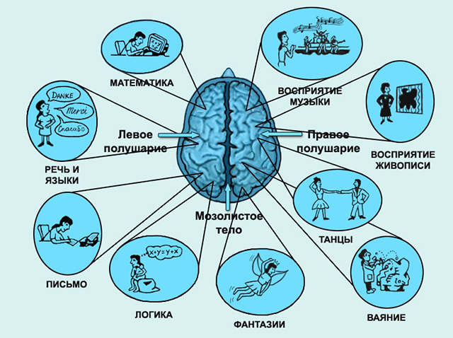
The main mass and volume of the brain fall precisely on the final section or the cerebral hemispheres. There are two hemispheres: the left one, which is mostly responsible for analytical thinking and speech functions of the body, and the right one, the main task of which is abstract thinking and all processes associated with creativity and interaction with the outside world.
The structure of the telencephalon
The cerebral hemispheres are the main "processing unit" of the CNS. Despite the different "specialization" these segments are complementary to each other.
The cerebral hemispheres are a complex system of interaction between the nuclei of nerve cells and nerve-conducting tissues connecting the main parts of the brain. The upper surface, called the cortex, is made up of a huge number of nerve cells. It's called gray matter. In the light of the general evolutionary development, the cortex is the youngest and most developed formation of the central nervous system and it has reached the highest development in humans. It is she who is responsible for the formation of higher neuropsychic functions and complex forms of human behavior. To increase the usable area, the surface of the hemispheres is assembled into folds or convolutions. The inner surface of the cerebral hemispheres consists of white matter - processes of nerve cells responsible for conducting nerve impulses and communication with the rest of the CNS segments.
In turn, each of the hemispheres is conditionally divided into 4 parts or lobes: occipital, parietal, temporal and frontal.
Occipital lobes
The main function of this conditional part is the processing of neural signals coming from the visual centers. It is here that the usual concepts of color, volume and other three-dimensional properties of a visible object are formed from light stimuli.

parietal lobes
This segment is responsible for the occurrence of pain sensations and the processing of signals from the body's thermal receptors. This is where their work ends.
The parietal lobe of the left hemisphere is responsible for structuring information packages, allows you to operate with logical operators, count and read. Also, this area forms awareness of the integral structure of the human body, the definition of the right and left parts, the coordination of individual movements into a single whole.
The right one is engaged in the generalization of information flows that are generated by the occipital lobes and the left parietal. On this site, a general three-dimensional picture of the perception of the environment, spatial position and orientation, miscalculation of perspective is formed.
temporal lobes
This segment can be compared with the "hard drive" of a computer - a long-term storage of information. It is here that all the memories and knowledge of a person collected over a lifetime are stored. The right temporal lobe is responsible for visual memory - the memory of images. Left - here all the concepts and descriptions of individual objects are stored, there is an interpretation and comparison of images, their names and characteristics.
As for speech recognition, both temporal lobes are involved in this procedure. However, their functions are different. If the left lobe is designed to recognize the semantic load of the words heard, then the right lobe interprets the intonation coloring and compares it with the speaker's facial expressions. Another function of this part of the brain is the perception and decoding of neural impulses coming from the olfactory receptors of the nose.
frontal lobes
This part is responsible for such properties of our consciousness as critical self-assessment, the adequacy of behavior, awareness of the degree of meaninglessness of actions, mood. The general behavior of a person also depends on the correct functioning of the frontal lobes of the brain, violations lead to inadequacy and asocial behavior. The process of learning, mastering skills, acquiring conditioned reflexes depends on the correct functioning of this part of the brain. This also applies to the degree of activity and curiosity of a person, his initiative and awareness of decisions.
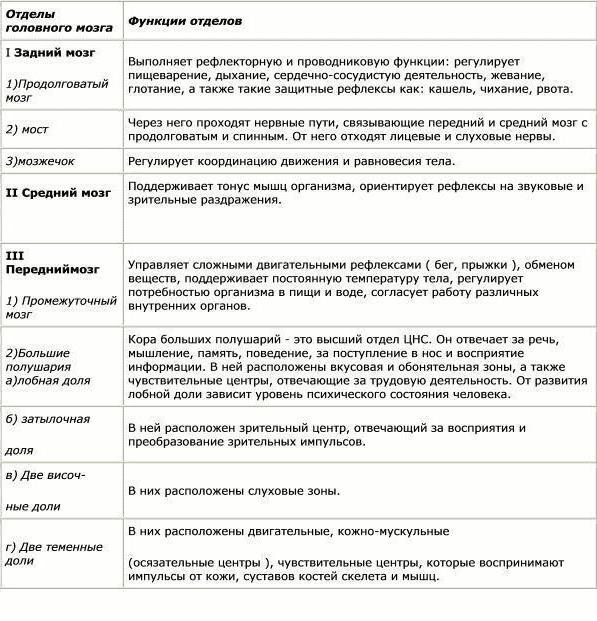
To systematize the functions of the GM, they are presented in the table:
| Department of the brain | Functions |
|---|---|
| Medulla | Control of basic protective reflexes. Control of unconscious reflexes. Control of balance and coordination of movements. |
| midbrain | Processing of nerve impulses, visual and auditory centers, response to them. Regulation of body temperature, muscle tone, arousal, sleep. |
| diencephalon Hypothalamus Epithalamus |
Secretion of hormones and regulation of the endocrine system of the body. Awareness of the surrounding world, processing and interpretation of impulses coming from peripheral receptors. Processing information from peripheral receptors Control of heart rate and blood pressure. Production of hormones. Control of hunger, thirst, satiety. Regulation of the daily biological rhythm, regulation of the body's metabolism. |
| Hind brain Cerebellum |
Coordination of motor functions. Regulation of cognitive functions: attention, understanding, awareness of language, regulation of the sensation of fear, sense of time, awareness of the nature of pleasure. |
| Large hemispheres of the brain Occipital lobes parietal lobes temporal lobes Frontal lobes. |
Processing of neural signals coming from the eyes. Interpretation of pain and heat sensations, responsibility for the ability to read and write, logical and analytical thinking ability. Long-term storage of information. Interpretation and comparison of information, recognition of speech and facial expressions, decoding of neural impulses coming from olfactory receptors. Critical self-assessment, adequacy of behavior, mood. The process of learning, mastering skills, acquiring conditioned reflexes. |
The interaction of brain regions
In addition to the fact that each part of the brain has its own tasks, the integral structure determines consciousness, character, temperament and other psychological features of behavior. The formation of certain types is determined by varying degrees of influence and activity of one or another segment of the brain.
The first psychotype or choleric. The formation of this type of temperament occurs with the dominant influence of the frontal lobes of the cortex and one of the subdivisions of the diencephalon - the hypothalamus. The first generates purposefulness and desire, the second section reinforces these emotions with the necessary hormones.
A characteristic interaction of the departments that determines the second type of temperament - sanguine, is the joint work of the hypothalamus and the hippocampus (the lower part of the temporal lobes). The main function of the hippocampus is to maintain short-term memory and convert acquired knowledge into long-term memory. The result of this interaction is an open, inquisitive and interested type of human behavior.
Melancholics are the third type of temperamental behavior. This option is formed with increased interaction between the hippocampus and another formation of the cerebral hemispheres - the amygdala. At the same time, the activity of the cortex and hypothalamus is reduced. The amygdala takes on the entire “blow” of excitatory signals. But since the perception of the main parts of the brain is inhibited, the response to excitation is low, which in turn affects behavior.
In turn, by forming strong connections, the frontal lobe is able to set an active model of behavior. When the cortex of this area interacts with the tonsils, the central nervous system generates only highly significant impulses, while ignoring insignificant events. All this leads to the formation of a Phlegmatic behavior model - a strong, purposeful person with an awareness of priority goals.
What is the carrier of consciousness - brain cells or electrical signals generated by them? Where do the consciousness and personality of a person come from and where do they go at the end of their journey? These questions concern many.
The human brain is one of the most mysterious organs of the human body. Scientists still cannot fully understand the mechanism of mental activity, the functioning of consciousness and subconsciousness.
Structure
In the course of evolution, a strong cranium has formed around the human brain, protecting this organ that is vulnerable to physical influences. The brain occupies more than 90% of the space of the skull. It consists of three main parts:- large hemispheres;
- brain stem;
- cerebellum.
It is also customary to distinguish five sections of the brain:
- forebrain (large hemispheres);
- hindbrain (cerebellum, pons Varolii);
- medulla;
- midbrain;
- intermediate brain.
Next comes Pons- This is a roller of nerve transverse fibers and gray matter. The main artery that feeds the brain passes through it. It starts above the medulla oblongata and passes into the cerebellum.
Cerebellum consists of two small hemispheres connected by a "worm", as well as white matter and gray matter covering it. This department is connected by pairs of "legs" to the oblong bridge, the cerebellum and the midbrain.
midbrain consists of two visual hillocks, and two auditory (quadrigemina). Nerve fibers that connect the brain with the spinal cord depart from these tubercles.
Large hemispheres of the brain separated by a deep fissure with the corpus callosum inside, which connects these two sections of the brain. Each hemisphere has a frontal, temporal, parietal and occipital. The hemispheres are covered by the cerebral cortex, in which all thought processes take place.
In addition, there are three layers of the brain:
- Hard, which is the periosteum of the inner surface of the skull. A large number of pain receptors are concentrated in this shell.
- Arachnoid, which is closely adjacent to the cerebral cortex, but does not line the gyrus. The space between it and the hard shell is filled with serous fluid, and the space between it and the cerebral cortex is filled with cerebrospinal fluid.
- Soft, consisting of a system of blood vessels and connective tissue, in contact with the entire surface of the substance of the brain, and nourishing it.
Functions and tasks
Our brain takes part in the processing of information coming from the entire set of receptors, controls the movements of the human body, and also carries out the highest function of the human body - thinking. Each part of the brain is responsible for performing certain functions.Medulla contains nerve centers that ensure the normal functioning of protective reflexes - sneezing, coughing, blinking, vomiting. He also "rules" the respiratory and swallowing reflexes, salivation and secretion of gastric juice.
Pons responsible for the normal movement of the eyeballs and the coordination of the facial muscles.
Cerebellum exercises control over the consistency and coordination of movement.
midbrain provides a regulatory function in relation to the acuity of hearing and clarity of vision. This part of the brain controls the expansion-constriction of the pupil, changes in the curvature of the lens of the eye, and is responsible for the muscle tone of the eye. It also contains the nerve centers of the orientation reflex in space.

diencephalon includes:
- thalamus- a kind of "switch" that processes and forms sensations from information from temperature, pain, vibration, muscle, taste, tactile, auditory, olfactory receptors, one of the subcortical visual centers. Also, this site is responsible for changing the states of sleep and wakefulness in the body.
- Hypothalamus- this small area performs the most important task of controlling heart rate, thermoregulation of the body, blood pressure. It also "manages" the mechanisms of emotional regulation - it affects the endocrine system in order to develop the hormones necessary to overcome stressful situations. The hypothalamus regulates hunger, thirst and satiety. It is the center of pleasure and sexuality.
- Pituitary- this brain appendage produces growth hormones of puberty, development and functioning.
- Epithalamus- includes the pineal gland, which regulates daily biological rhythms, releasing hormones at night for normal and long falling asleep, and during the day - for a normal mode of wakefulness and activity. Directly with the regulation of sleep and wakefulness is associated with the control of the body's adaptation to lighting conditions. The pineal gland is able to pick up vibrations of light waves even through the cranium, and respond to them by releasing the necessary hormones. Also, this small part of the brain regulates the rate of metabolism in the body (metabolism).
Left cerebral hemisphere- exercises control over the speech functions of the body, the implementation of analytical activities, mathematical calculations. Here abstract thinking is formed, the movement of the left limbs is controlled.
Each of the hemispheres of the brain is divided into 4 lobes:
1. Frontal lobes- they can be compared with the navigational cabin of the ship. They ensure the maintenance of the vertical position of the human body. Also, this site is responsible for how active and inquisitive a person is, initiative and independent in making decisions.
In the frontal lobes, processes of critical self-evaluation take place. Any violations in the frontal lobes lead to the manifestation of inadequacy in behavior, senselessness of actions, apathy and sudden mood swings. Also, "logging" manages human behavior and control over it - the prevention of deviations, socially unacceptable actions.

Actions of an arbitrary nature, their planning, mastery of skills and abilities also depend on the frontal lobes. Here, frequently repeated actions are brought to automatism.
In the left (dominant) lobe, control is exercised over human speech, ensuring abstract thinking.
2. Temporal lobes- this is a long-term storage. The left (dominant) share stores information about the specific names of objects, the links between them. The right lobe is responsible for visual memory and imagery.
Their important function is also speech recognition. The left lobe deciphers for consciousness the semantic load of the spoken words, and the right lobe provides an understanding of their intonational coloring and facial expressions, explaining the mood of the speaker and the degree of his goodwill towards us.
The temporal lobes also provide the perception of olfactory information.
3. Parietal lobes- participate in the perception of pain, feelings of cold, heat. The functions of the right and left lobes are different.
The left (dominant) share provides the processes of synthesizing information fragments, combining them into a single system, allows a person to read and count. This share is responsible for the assimilation of a certain algorithm of movements leading to a specific result, the feeling of individual parts of one's own body and a sense of its integrity, the definition of the right and left sides.
The right (non-dominant) lobe transforms the entire set of information coming from the occipital lobes, forming a three-dimensional picture of the world, provides orientation in space, determining the distance between objects and to them.
4. Occipital lobes- processing visual information. perceive objects of the surrounding world as a set of stimuli that reflect light on the retina in different ways. The occipital lobes convert light signals into information about the color, movement and shape of objects that are understandable to the parietal lobes, which form three-dimensional images in our minds.
Brain diseases
The list of brain diseases is quite large, we will give the most common and dangerous of them.Conventionally, they can be divided into:
- tumor;
- viral;
- vascular;
- neurodegenerative.

Tumor diseases. The number of brain tumors is very diverse. They can be malignant or benign. Tumors arise as a result of a failure in cell reproduction, when cells must die and give way to others. Instead, they multiply uncontrollably and rapidly, crowding out healthy tissue.
Symptoms may include: nausea,
The brain is the main regulator of the functions of any living organism, one of the elements Until now, medical scientists have been studying the features of the brain and discovering new incredible possibilities. This is a very complex organ that connects our body with the external environment. The parts of the brain and their functions regulate all life processes. External receptors catch signals and inform any part of the brain about incoming stimuli (light, sound, tactile, and many others). The response is immediate. Let's take a closer look at how our head "processor" works.
General description of the brain
The parts of the brain and their functions completely control our life processes. The human brain consists of 25 billion neurons. This incredible number of cells forms the gray matter. The brain covers several layers:
- soft;
- hard;
- arachnoid (cerebrospinal fluid circulates here).
Liquor is a cerebrospinal fluid, in the brain it plays the role of a shock absorber, a protector from any impact force.
In both men and women, the brain is developed in exactly the same way, although its weight is different. More recently, the debate has subsided that the weight of the brain plays some role in mental development and intellectual abilities. The conclusion is unambiguous - it is not. The weight of the brain is approximately 2% of the total mass of a person. In men, its average weight is 1,370 g, and in women - 1,240 g. The functions of the parts of the human brain are developed in a standard way, vital activity depends on them. Mental abilities depend on the quantitative connections created in the brain. Each brain cell is a neuron that generates and transmits impulses.
The cavities inside the brain are called ventricles. The cranial paired nerves go to different departments.
Functions of the brain regions (table)
Every part of the brain has a job to do. The table below clearly demonstrates this. The brain, like a computer, clearly performs its tasks, receiving commands from the outside world.
The functions of the brain regions, the table reveals schematically and succinctly.
Let's take a closer look at the parts of the brain below.
Structure
The picture shows how the brain works. Despite this, the most significant part is occupied by all parts of the brain and their functions play a huge role in the functioning of the body. There are five main divisions:
- final (of the total mass is 80%);
- posterior (bridge and cerebellum);
- intermediate;
- oblong;
- average.
At the same time, the brain is divided into three main parts: the brain stem, the cerebellum, and the two cerebral hemispheres.

telencephalon
It is impossible to briefly describe the structure of the brain. To understand the parts of the brain and their functions, it is necessary to study their structure closely.
The telencephalon stretches from the frontal to the occipital bone. Two cerebral hemispheres are considered here: left and right. This department differs from others in the largest number of furrows and convolutions. The development and structure of the brain are closely linked. Experts have identified three types of bark:
- ancient (with olfactory tubercle, anterior perforated substance, semilunar subcallosal and lateral subcallosal gyrus);
- old (with dentate gyrus - fascia and hippocampus);
- new (represents the rest of the cortex).
The hemispheres are separated by a longitudinal groove, in its depths there is a vault and a corpus callosum, which connect the hemispheres. The corpus callosum itself is lined and belongs to the neocortex. The structure of the hemispheres is quite complex and resembles a multi-level system. Here, the frontal, temporal, parietal and occipital lobes, subcortex and cortex are distinguished. The large hemispheres perform a huge number of functions. It is worth noting that the left hemisphere commands the right side of the body, and the right, on the contrary, the left.
Bark
The surface layer of the brain is the cortex, it has a thickness of 3 mm, covers the hemispheres. The structure consists of vertical nerve cells with processes. The cortex also contains efferent and afferent nerve fibers, as well as neuroglia. The parts of the brain and their functions are discussed in the table, but what is the cortex? Its complex structure has horizontal layering. The building has six layers:
- external pyramidal;
- external granular;
- internal granular;
- molecular;
- internal pyramidal;
- with spindle cells.
Each has a different width, density, shape of neurons. Vertical bundles of nerve fibers give the cortex a vertical striation. The area of the cortex is approximately 2,200 square centimeters, the number of neurons here reaches ten billion.
Parts of the brain and their functions: cortex
The cortex controls several specific bodily functions. Each share is responsible for its own parameters. Let's take a closer look at the functions associated with hotels:
- temporal - controls the sense of smell and hearing;
- parietal - responsible for taste and touch;
- occipital - vision;
- frontal - complex thinking, movement and speech.
Each neuron contacts other neurons, there are up to ten thousand contacts (gray matter). Nerve fibers are white matter. Some part unites the hemispheres of the brain. White matter includes three types of fibers:
- association links connect different cortical areas in one hemisphere;
- commissural connect the hemispheres to each other;
- projection ones communicate with lower formations, have paths of analyzers.
Considering the structure and functions of the brain, it is necessary to emphasize the role of gray and white matter. The hemispheres inside have (gray matter), their main function is the transmission of information. The white matter is located between the cerebral cortex and the basal ganglia. There are four parts here:
- between furrows in convolutions;
- in the outer places of the hemispheres;
- included in the inner capsule;
- located in the corpus callosum.
The white matter located here is formed by nerve fibers and connects the cortex of the convolutions with the underlying sections. form the subcortex of the brain.
The telencephalon - manages all the vital functions of the body, as well as the intellectual abilities of a person.

diencephalon
The brain regions and their functions (table above) include the diencephalon. If you look in more detail, it is worth saying that it consists of ventral and dorsal parts. The hypothalamus belongs to the ventral, and the thalamus, metathalamus, and epithalamus to the dorsal.
The thalamus is a mediator that directs the received irritations to the hemispheres. It is often referred to as the "optic tubercle". It helps the body quickly adapt to changes in the external environment. The thalamus is connected to the cerebellum via the limbic system.
The hypothalamus controls autonomic functions. The influence goes through the nervous system, and, of course, the endocrine glands. Regulates the work of the endocrine glands, controls metabolism. The pituitary gland is located directly below it. Body temperature, cardiovascular and digestive systems are regulated. The hypothalamus also controls our eating and drinking behavior, regulates wakefulness and sleep.
Rear
The hindbrain includes the pons located in front and the cerebellum, which is located behind. Studying the structure and functions of the brain regions, let's take a closer look at the structure of the bridge: the dorsal surface is covered by the cerebellum, the ventral one is represented by a fibrous structure. The fibers are directed transversely in this section. On each side of the bridge, they depart to the cerebellar middle peduncle. In appearance, the bridge resembles a thickened white roller located above the medulla oblongata. The nerve roots exit into the bulbar pontine groove.
The structure of the posterior bridge: on the frontal section, it can be seen that there is a section of the anterior (large ventral) and posterior (small dorsal) parts. Between them, the trapezoid body serves as a boundary, the transverse thick fibers of which are considered to be the auditory pathway. Conductor function is completely dependent on the hindbrain.
Cerebellum (small brain)
The table "Department of the brain, structure, functions" indicates that the cerebellum is responsible for the coordination and movement of the body. This department is located behind the bridge. The cerebellum is often referred to as the "small brain". It occupies the posterior cranial fossa, covers the rhomboid. The mass of the cerebellum ranges from 130 to 160 g. Above are the large hemispheres, which are separated by a transverse fissure. The lower part of the cerebellum is adjacent to the medulla oblongata.
Two hemispheres are distinguished here, the lower, upper surface and the worm. The boundary between them is called a horizontal deep slit. A lot of cracks cut the surface of the cerebellum, between them there are thin convolutions (rollers). Between the grooves there are groups of convolutions, divided into lobules, they represent the lobes of the cerebellum (posterior, flocculent-nodular, anterior).
The cerebellum contains both grays, and grays are located on the periphery, forming a cortex with molecular and pear-shaped neurons, and a granular layer. Under the cortex there is a white substance that penetrates into the gyrus. In the white matter there are blotches of gray (its nuclei). In cross section, this ratio is similar to a tree. Those who know the structure of the human brain, the functions of its departments, will easily answer that the cerebellum is the regulator of the coordination of the movements of our body.

midbrain
The midbrain is located in the region of the anterior pons and goes to the papillary bodies, as well as to the optic tracts. Here clusters of nuclei are distinguished, which are called tubercles of the quadrigemina. The structure and functions of the brain regions (table) indicate that this department is responsible for latent vision, the orienting reflex, gives orientation to reflexes to visual and sound stimuli, and also maintains muscle tone in the human body.
medulla oblongata: brainstem
The medulla oblongata is a natural extension of the spinal cord. That is why the structure has a lot in common. This becomes especially clear if we examine the white matter in detail. It is represented by short and long nerve fibers. In the form of nuclei, gray matter is represented here. Parts of the brain and their functions (the table is presented above) indicates that the medulla oblongata controls our balance, coordination, regulates metabolism, controls breathing and blood circulation. It is also responsible for such important reflexes of our body as sneezing and coughing, vomiting.
The brain stem is divided into the hindbrain and midbrain. The trunk is called the middle, oblong, bridge and diencephalon. Its structure is descending and ascending paths connecting the trunk with the spinal cord and brain. In this part, control over the heartbeat, breathing, articulate speech is carried out.
The brain, together with the membranes covering it, occupies the entire cavity of the skull. Its mass in an adult is on average 1360-1375 g. In a newborn, the mass of the brain is 370-400 g. During the first year of a child's life, it doubles, and by the age of 6 it increases 3 times. Then there is a slow addition of brain mass, which ends at the age of 20-25.
Sections of the brain
In accordance with the five cerebral vesicles from which the brain developed, five main sections are distinguished in it:
1. medulla;
2. hindbrain, consisting of bridge and cerebellum;
3. midbrain, including two legs of the brain and the roof of the midbrain with two pairs of mounds;
4. diencephalon, the main formations of which are two thalamus, with two pairs of geniculate bodies, and the hypothalamus;
5. telencephalon, represented by two hemispheres.
1. Medulla oblongata is a continuation of the spinal cord. It contains the nuclei of VIII-XII pairs of cranial nerves. Here are vital centers for the regulation of respiration, cardiovascular activity, digestion, and metabolism. The nuclei of the medulla oblongata are involved in the implementation of unconditioned food reflexes (separation of digestive juices, sucking, swallowing), protective reflexes (vomiting, sneezing, coughing, blinking). The conductor function of the medulla oblongata is to transmit impulses from the spinal cord to the brain and vice versa.
2. Cerebellum and pons form hindbrain. Nerve pathways pass through the bridge, connecting the forebrain and midbrain with the medulla oblongata and spinal cord. The nuclei of the V-VIII pairs of cranial nerves are located in the bridge. The gray matter of the cerebellum is outside and forms a cortex with a layer of 1-2.5 mm. The cerebellum is formed by two hemispheres connected by a worm. The nuclei of the cerebellum provide coordination of complex motor acts of the body. The cerebral hemispheres through the cerebellum regulate skeletal muscle tone and coordinate body movements. The cerebellum takes part in the regulation of some autonomic functions (blood composition, vascular reflexes).
3.Midbrain located between the pons and diencephalon. Consists of quadrigemina and legs of the brain . Through the midbrain, ascending paths pass to the cerebral cortex and cerebellum and descending paths to the medulla oblongata and spinal cord (conductor function). The midbrain contains the nuclei of the III and IV pairs of cranial nerves. With their participation, primary orienting reflexes to light and sound are carried out: eye movement, head turning towards the source of irritation. The midbrain is also involved in maintaining skeletal muscle tone.
4. Diencephalon located above the midbrain. Its main divisions are thalamus (optical tubercles) and hypothalamus (subtuberous area). Centripetal impulses from all receptors of the body (with the exception of the olfactory one) pass through the thalamus to the cerebral cortex. Information receives the corresponding emotional coloring in the thalamus and is transmitted to the cerebral hemispheres. The hypothalamus is the main subcortical center for the regulation of the autonomic functions of the body, all types of metabolism, body temperature, the constancy of the internal environment (homeostasis), and the activity of the endocrine system. The hypothalamus contains the centers of satiety, hunger, thirst, and pleasure. The nuclei of the hypothalamus are involved in the regulation of the alternation of sleep and wakefulness (pineal gland).
Ventricles The brain is a system of cavities. They contain cerebrospinal fluid.
- Lateral ventricles are cavities in the brain that contain cerebrospinal fluid. Such ventricles are the largest in the ventricular system. The left ventricle is called the first, and the right - the second. It is worth noting that the lateral ventricles communicate with the third ventricle using the interventricular or Monroe foramina. Their location is below the corpus callosum, on both sides of the midline, symmetrically. Each lateral ventricle has an anterior horn, posterior horn, body, and inferior horn.
- third ventricle- located between the visual tubercles. It has an annular shape, since intermediate visual tubercles grow into it. The walls of the ventricle are filled with central gray medulla. It contains subcortical vegetative centers. The third ventricle communicates with the aqueduct of the midbrain. Behind the nasal commissure, it communicates through the interventricular foramen with the lateral ventricles of the brain.
- fourth ventricle-located between the medulla oblongata and the cerebellum. The arch of this ventricle is the cerebral sails and the worm, and the bottom is the bridge and the medulla oblongata.
5. Forebrain- the largest and most developed part of the brain. It is represented by two hemispheres - left and right, separated by a longitudinal slit. The hemispheres are connected by a thick horizontal plate - corpus callosum, which is formed by nerve fibers running transversely from one hemisphere to the other. Three furrows - central, parietal-occipital and lateral - divide each hemisphere into four lobes: frontal, parietal, temporal and occipital. The fifth - the insular lobe (islet) - is embedded in the depths of the lateral fossa of the large brain, which separates the frontal lobe from the temporal lobe.
Outside the hemisphere covers a layer of gray matter - bark, located inside white matter and subcortical nuclei. The subcortical nuclei are a phylogenetically ancient part of the brain that controls unconscious automatic actions (instinctive behavior). The white matter of the forebrain is formed by nerve fibers that connect different parts of the brain.
Bark brain has a thickness of 1.3-4.5 mm. Due to the presence of folds, convolutions and furrows, the total area of the cortex of an adult is 2000-2500 cm 2. The cortex consists of 12-18 billion nerve cells arranged in six layers.
Cells are classified according to morphological features into the main types: pyramidal, spindle-shaped, stellate, granular. Functionally, neurons are divided into sensory, motor and intermediate (intercalary). Pyramidal and fusiform cells perform an efferent function, and stellate cells perform an afferent one.
Layered organization of the neocortex:
I. Molecular. This layer contains many fibers forming a dense plexus parallel to the surface, but few cells.
II. External granular. It densely contains small neurons of various shapes, among which are small pyramidal cells. Nerve fibers here are oriented mainly parallel to the surface of the cortex.
III. External pyramidal. It consists mainly of pyramidal neurons.
IV. Internal granular. In this layer, small neurons of various sizes (stellate cells) are diffusely located, between which dense bundles of fibers parallel to the surface of the cortex pass.
V. Internal pyramidal. It consists mainly of medium and large pyramidal cells; for example, Betz's giant pyramidal cells in the precentral gyrus.
VI. A layer of spindle cells. Here are predominantly spindle-shaped neurons. The deep part of this layer passes into the white matter of the brain.
Although the cerebral cortex functions as a whole, the functions of its individual sections are not the same. AT sensory (sensitive) zones the cortex receives impulses from all receptors of the body. So, the visual zone of the cortex is located in the occipital lobe, the auditory - in the temporal, etc. association areas the cortex stores, evaluates, compares incoming information with previously received information, etc. Thus, the processes of memorization, learning, and thinking take place in this zone. Motor (motor) zones are responsible for conscious movements. From them, nerve impulses are sent to the striated muscles.
1 - corpus callosum;
2 - vault;
3 - thalamus;
4 - roof of the midbrain;
5 - mastoid body;
6 - aqueduct of the midbrain;
7 - leg of the brain;
8 - optic chiasm;
9 - IV ventricle;
10 - pituitary gland;
11 - bridge;
12 - cerebellum
The brain has a complex structure and is the central organ of the nervous system. Parts of the brain interact with each other through neural connections that regulate the activity of the whole organism.
The human nervous system has been studied quite well, which made it possible to describe in detail what departments the brain consists of and their relationship with various organs, as well as the impact on behavioral responses. The CNS organ contains billions of neurons through which electrical impulses pass, transmitting information to brain cells from internal organs and systems.
Brain structures are firmly protected from the effects of negative external factors:
- Cerebrospinal fluid (CSF) - is located between the membranes and the surface of the organ. The cerebrospinal fluid acts as a shock absorber, protecting structures from damage and friction. The fluid continuously circulates in the ventricles of the brain, in the subarachnoid space and the spinal canal. In addition to mechanical protection, it also maintains stable intracranial pressure and metabolic processes;
- Arachnoid membrane (arachnoid) - the middle shell, the deepest and softest. It is formed from connective tissue and contains a large amount of collagen fibers. Participates in the exchange of cerebrospinal fluid. The arachnoid membrane contains very thin thread-like strands that are woven into the soft shell;
- The inner shell (soft) - tightly adheres to the structures, filling all spaces (crevices, furrows). Consists of a loose connective tissue permeated with a circulatory network that delivers nutrients to the cells of the body;
- Superficial shell (solid) - formed from dense connective tissue and has two surfaces. The outer surface contains a large number of vessels and has a rough surface. The inner surface is smooth and adheres tightly to the bones - fuses with the periosteum of the cranium and the sutures of the fornix;
- The cranium - forms a protective frame for the structures of the brain and its membranes, consists of 23 bones connected to each other. The skull serves as an attachment site for the soft tissues of the brain.
The cells of the brain structures are formed from the bodies of neurons (gray matter, the main component of the nervous system) and the myelin sheath (white matter). Each functionally active cell of an organ has a long process (axon) that branches and connects to another neuron (synapse).
Thus, a kind of circuit is obtained for transmitting and receiving an electrical impulse from one neuron to another. Signals to brain structures come through the spinal cord and cranial nerves extending from the trunk. In some parts of the brain, neurons are transformed by synthesizing hormones.
The human brain consists of: anterior, middle and posterior sections. The scientific works of researchers describe the brain after opening the cranium as two large hemispheres and an extended formation (trunk), so the brain is usually divided into three sections. The hemispheres are separated by a longitudinal groove - an interlacing of nerve fibers (corpus callosum), which looks like a wide strip, consists of axons.
The functions of these parts of the brain are in the formation of thought processes and the possibility of sensory perception. Each hemisphere has a different functionality and is responsible for the opposite half of the body (left for the right half and vice versa). The main parts of the brain are formed by dividing the organ with the help of furrows and convolutions.
The structures of the brain are divided into 5 sections:
- Hind brain (rhomboid);
- Average;
- Front;
- Finite;
- Olfactory.
The organ of the central nervous system has a high plasticity - if one of the departments is damaged, compensatory capabilities are temporarily triggered, allowing it to perform the functions of the disturbed department. Conventionally, the brain is divided into: the right hemisphere and the left hemisphere, the cerebellum, the medulla oblongata. These three departments are connected in a single network, but differ in functionality.
The cerebral cortex
The cortex of the hemispheres forms a thin layer of gray matter responsible for higher mental function. On the surface of the cortex, furrows can be visually seen, which is why all parts of the brain have a folded surface. The central organ of each person has a different shape of furrows, depth and length, thus, an individual pattern is formed.

Studies of brain structures made it possible to determine the most ancient cortical layer and the evolutionary development of the organ by histological analysis. The bark is divided into several types:
- Archipallium - the oldest part of the cortex, regulates emotions and instincts;
- The paleopallium is the younger part of the cortex, is responsible for vegetative regulation and maintains the physiological balance of the whole organism;
- Neocortex - a new area of the cortex, forms the upper layer of the cerebral hemispheres;
- Mesocortex - consists of an intermediate old and new cortex.
All areas of the cortex are in close interaction with each other, as well as with subcortical structures. The subcortex includes the following structures:
- The thalamus (visual tubercles) is an accumulation of a large mass of gray matter. The thalamus contains sensory and motor nuclei, nerve fibers allow it to be connected to many parts of the cortex. The visual hillocks are connected to the limbic system (hippocampus) and are involved in the formation of emotions and spatial memory;
- Basal ganglia (nucleus) - accumulation of white matter in the thickness of gray. The layer is located on the side of the thalamus, near the base of the hemispheres. The basal nuclei carry out the higher processes of nervous activity, the active phase of work occurs during the daytime, and stops during sleep. Neurons in the nuclei are activated during the mental work of the organ (concentration of attention), and produce electrochemical impulses;
- The nuclei of the brain stem - regulate the mechanisms of redistribution of muscle tone, and are responsible for maintaining balance;
- Spinal cord - located in the spinal canal, and has a cavity filled with CSF. It is presented in the form of a long cord and provides a connection between the large brain and the periphery. The spinal cord is divided into segments and performs reflex activity. Information flows through the spinal canal to the brain.
The hierarchy of these structures in relation to the cortex is lower, but each performs important functions and, in case of violations, independent self-government is launched. The subcortical region is represented by a complex of various formations that are involved in the regulation of behavioral responses.
Lobes of the brain and centers
The mass of the central organ is about 2% of the total weight of a person. Each cell of the body needs an active blood supply and consumes up to 15% of the total volume of circulating blood in the body. The blood supply to the brain tissues is a separate functional system - it supports the vital activity of each cell, delivering nutrients and oxygen (consuming 20% of the total volume).

Arteries form a vicious circle, with the activity of neurons, blood flow to this area also increases. Blood and brain tissues are delimited from each other by a physiological barrier (blood-brain barrier) - it provides selective permeability of substances, protecting the main sections of the organ from various infections. The outflow of blood from the central nervous system is carried out through the jugular veins.
The left and right hemisphere includes five departments:
- The frontal lobe is the most massive part of the hemispheres; damage to this area results in loss of behavioral control. The frontal pole is responsible for coordination of movements and speech skills;
- Parietal lobe - responsible for the analysis of various sensations, including body perception and the development of various skills (reading, counting);
- Occipital lobe - this part processes incoming optical signals, creating visual images;
- Temporal lobe - processes incoming audio signals. Each sound is analyzed for correct perception. This part of the brain is also responsible for the emotional background, which is reflected in facial reactions. The temporal lobes are the center for storing incoming information (long-term memory);
- Insular - divides the frontal and temporal parts, this lobe is responsible for consciousness (reaction to various situations). The insular lobe processes all signals from the senses, forming images.
Each hemisphere has protrusions, which are called the pole:
- Frontal - in front;
- Occipital - behind;
- Side - temporal.
The hemispheres also have three surfaces: convexital - convex, inferior and medial. Each surface passes from one to another, forming edges (superior, inferolateral, inferomedial). What each part of the brain is responsible for and what functions it performs depends on the centers located in them. Violation of the vital center entails a serious consequence - a fatal outcome.
In which part of the brain are the centers of human speech and other active areas in the cortical structure, depends on the anatomical division of the cerebral hemispheres, with the help of furrows. The formation of furrows is a process of evolutionary development of the organ, since the growth of the final brain structures is limited by the cranium. Intensive tissue growth led to the ingrowth of gray matter into the thickness of the white.
frontal lobe
The frontal part is formed by the cerebral cortex and is separated from the other lobes by furrows. The central sulcus delimits the frontal-parietal part, and the lateral sulcus delimits from the temporal region. This part by volume makes up a third of the entire mass of the cortex and is divided into various fields (centers) that are responsible for a particular system or skill.
Functions of the frontal lobe and centers:
- Information processing center and expression of emotions;
- Center for the motor organization of speech (Broca's area);
- Sensory speech zone (Wernicke) - responsible for the process of assimilation of the received information and understanding of written and oral speech;
- Head and eye rotation analyzer;
- thought processes;
- Regulation of conscious behavior;
- Movement coordination.
The size of the fields refers to the individual characteristics of a person and depends on the activity of neurons. The central gyrus in the frontal zone is divided into three parts, and each of them regulates the physical activity of the muscles in a certain area (facial expressions, motor activity of the upper and lower extremities, the human body).
parietal lobe
The parietal part is formed by the cerebral cortex and is separated from other zones by a central sulcus. The parieto-occipital sulcus (posteriorly) extends to the temporal sulcus. Nerve fibers depart from the parietal zone, connecting the entire part with muscle fibers and receptors.
Functions of the parietal zone and centers:
- Computing center;
- Body thermoregulation center;
- Spatial analysis;
- Sensory center (response to sensations);
- Responsible for complex motor skills;
- Center for Visual Analysis of Written Speech.
The left part of the parietal zone is involved in the impulse to motor acts. The development of furrows and convolutions in this area is directly related to the conduction of nerve impulses. The parietal region allows, without the participation of visual analyzers, to determine the location of any part of the body or to indicate the shape of an object and its size.
The temporal region is formed by the cortex of the hemispheres, the lateral groove delimits the lobe from the parietal and frontal regions. The share has two furrows and four convolutions, interacts with the limbic system. The main sulci form three convolutions, dividing the temporal part into small sections (upper, middle, lower).

In the depth of the lateral sulcus is the Geschl gyrus (a group of small convolutions). This area of the cortex has the clearest border lines. The upper part of the temple has a convex surface, and the lower part is concave.
The general functions of the temporal lobe are visual and auditory processing and language comprehension. Features of this area are expressed in a different functional orientation of the right temporal lobe and the left.
The work of the right lobe is more focused on the analysis of various emotions and their comparison with the facial expression of the interlocutor.
insular lobe
The islet is part of the cortical structure of the hemispheres and is located deep in the Sylvian furrow. This part is hidden under the frontal, parietal and temporal region. Visually resembles an inverted pyramid, where the base is facing the frontal part.

The perimeter of the insula is delimited by periinsular sulci, the central sulcus divides the entire lobe into two parts (larger - anterior, smaller - posterior). The anterior part contains short convolutions, and the posterior part contains two long ones.
The islet as a full-fledged share of the organ has been recognized only since 1888. Previously, the hemispheres were divided into four lobes, and the islet was considered only as a small formation. The insula connects the limbic system and the cerebral hemispheres.
The islet consists of several layers of neurons (from 3 to 5) that process sensory impulses and exercise sympathetic control of the cardiovascular system.
Functions of the insular lobe:
- Behavioral reactions and reciprocal emotions;
- Performs voluntary swallowing;
- Phonetic speech planning;
- Controls sympathetic and parasympathetic regulation.
The insular lobe supports subjective sensations that come from the internal organs in the form of signals (thirst, cold) and allows you to consciously perceive your own existence.
Functions of the main departments
Each of the five main departments performs different functions in the body and supports vital processes.
Correspondence between the functions and parts of the human brain:
| brain department | Functions performed |
|---|---|
| Rear | Responsible for the coordination of movements. |
| Front | Responsible for the intellectual capabilities of a person, the ability to analyze and store the information received. |
| Average | Responsible for physiological functions (vision, hearing, regulation of biorhythms and pain sensations). |
| Finite | Responsible for speech skills and vision. Controls skin-muscle sensitivity and the occurrence of conditioned reflexes. |
| Olfactory | Responsible for the function of various feelings in humans. |
The table reflects the overall functionality, the structure of each department in the central body, includes various structures and areas that are responsible for a specific function.
All parts of the brain work in conjunction with each other - this allows you to perform higher mental activity, through the reception and processing of information coming from the senses.
The posterior section of the central organ of the central nervous system includes the bulb (medulla oblongata), which is included in the stem part. The bulb is responsible for coordinating movements and maintaining balance in an upright position.
Anatomically, the structure is located between the exit of the first spinal nerve (occipital foramen area) and the pons (superior border). This department regulates the respiratory center - a vital department, if it is damaged, instant death occurs.
The main functions of the medulla oblongata:
- Regulation of blood circulation (work of the heart muscle, stabilization of blood pressure);
- Regulation of the digestive system (production of digestive enzymes, salivation);
- Regulation of muscle tone (rectifying, postural and labyrinth reflexes);
- Control of unconditioned reflexes (sneezing, vomiting, blinking, swallowing);
- Regulation of the respiratory center (state of the lung tissue and its stretching, gas composition).
The medulla oblongata has an internal and external structure. On the outer surface is the median line, which divides the pyramids (the connection of the cortex with the nuclei of cranial nerves and motor horns).
In the line there is a crossing of the nerve fiber and a corticospinal path is formed. To the side of the pyramid is an olive (oval extension). The pyramidal system allows a person to perform complex coordination of movements.
Internal structure (kernels of gray matter):
- Olive kernel (plate of gray matter);
- Nerve cells with complex connections (reticular formation);
- Nuclei of cranial nerves (glossopharyngeal, hypoglossal, accessory and vagus);
- The connection between the vital centers and the nucleus of the vagus nerve.
The bundles of axons in the bulb provide a connection between the spinal cord and other parts of the central nervous system (the pathways are long and short). Autonomic functions are regulated in the medulla oblongata.

The vasomotor center and nuclei of the vagus nerve invert the signals necessary to maintain tone - the arteries and arterioles are always slightly narrowed, and the activity of the heart is slowed down. In the bulb there are active poles that stimulate the production of various secrets: salivary, lacrimal, gastric enzymes, bile formation, pancreatic enzymes.
midbrain
The middle part of the body performs a lot of physiologically significant functions.
Anatomical structure:
- Four hills (two upper and two lower) - these hillocks form the upper surface of the middle part of the organ;
- Sylvius aqueduct - is a cavity;
- The cerebral peduncles are paired parts that connect to the tegmentum of the midbrain.
This department belongs to the stem structure of the organ and has a complex structure, despite its small size. The midbrain is the subcortical part of the brain, which is part of the motor center of the extrapyramidal system.
Functions of the inner brain:
- Responsible for vision;
- Controls movement;
- Regulates biorhythms (sleep and wakefulness);
- Responsible for concentration;
- Regulates pain;
- Responsible for hearing
- Regulates protective reflexes;
- Supports thermoregulation in the body.
In the thickness of the legs of the brain there are nerve fibers that concentrate almost all the paths of general sensitivity. Various lesions of the internal structure of the organ lead to impaired vision and hearing. Movement of the eyeballs becomes impossible, marked strabismus is noted together with hearing loss (bilateral). Often there are hallucinations, both auditory and visual.
Posterior, including cerebellum and pons
The hindbrain itself consists of the pons and the cerebellum, which are part of the rhomboid region. The cavity of the hindbrain communicates with the oblong (fourth ventricle). The pons varolii is located under the cerebellum and contains a large amount of nerve fiber, forming descending pathways that transmit information from the spinal cord to various parts of the head structures. The scheme of the bridge is presented in the form of a roller with a recess (basilar sulcus).

The third department of the central organ regulates the vestibular apparatus and the coordination of movements. These functions are provided by the cerebellum, which is also involved in the adaptation of the motor center in various disorders. The cerebellum is often called the small brain - this is due to the visual similarity with the main organ. The small brain is located in the cranial fossa and is protected by a hard shell.
Anatomical structure:
- Right hemisphere;
- Left hemisphere;
- Worm;
- Brain body.
The cerebellar hemispheres have a convex surface (lower), the upper part is flat. There is a gap on the posterior surface of the edges, the anterior edge with pronounced furrows. The lobules of the cerebellum on the surface are formed by small furrows and sheets, covered with bark from above.
The lobules are interconnected by a worm, from the cerebrum, the small one separates the gap, which includes the process of the dura mater (the cerebellum is stretched over the cranial fossa).
Legs depart from the cerebellum:
- The lower ones - to the medulla oblongata (nerve fibers coming from the spinal cord pass through the lower legs);
- Medium - to the bridge;
- Upper - to the midbrain.
Outside, the brain is covered by a layer of gray matter, under which are bundles of axons. If this area is damaged or anomalies in development, the muscles become atonic, a staggering gait and tremor of the limbs appear. There is also a change in handwriting.
The defeat of the pyramidal tracts located in the bridge leads to spastic paresis - a violation of facial expressions is associated with damage to this part of the brain.
diencephalon
This department is part of the front of the body and manages and switches all incoming information. The functions of the forebrain are the adaptive capabilities of the human body (external negative factors) and the regulation of the autonomic nervous system.

The diencephalon includes:
- thalamic region;
- Hypothalamic-pituitary system (hypothalamus and posterior pituitary gland);
- Epithalamus.
The hypothalamus regulates the work of internal organs and systems and is the center of pleasure. This part is represented as a small cluster of neurons that transmit signals to the pituitary gland.
The thalamus processes all signals coming from sensory receptors, redistributing them to the corresponding parts of the CNS organ.
The epithalamus synthesizes the hormone melatonin, which is involved in the regulation of biorhythms and the emotional background of a person.
The hypothalamus is part of an important system of the central nervous system - the limbic. This system performs a motivational-emotional function (adapts when habitual conditions change). The system is closely related to memory and sense of smell, evoking clear memories of a bright event or reproducing a pleasant smell (of food, perfume).
telencephalon
The youngest part of the brain is the terminal part. It is a fairly massive department of the central nervous system and is the most developed.
The telencephalon covers all departments and consists of:
- cerebral hemispheres;
- Nerve fiber plexus (corpus callosum);
- Alternating bands of gray and white matter (striatum);
- Structures associated with the sense of smell (olfactory brain).
In the cavity of the end part of the organ there are lateral ventricles, represented in each hemisphere (conditionally considered right and left).
End department functions:
- traffic regulation;
- Reproduction of sounds (speech);
- skin sensitivity;
- Auditory and gustatory sensations, smell.
The longitudinal fissure separates the left and right hemispheres, the corpus callosum (white matter plate) is located deep in the fissure. In the thickness of the white matter are the basal nuclei, which are responsible for the transfer of information from one department to another and perform basic functions.
The hemispheres control and are responsible for the work of the opposite side of the body (right for the left half and vice versa). The left hemisphere of the brain is responsible for memory, thought processes and individual talents in humans.
The right hemisphere in the brain is responsible for the processing of various information and imagination, which is also generated in dreams. All parts of the brain and the functions they perform are the joint work of the two hemispheres and the cortical part.
In each person, one part of the organ dominates, either the right or the left - which hemisphere is more active depends on individual characteristics.
Consistency of all structures of the brain allows you to perform all functions harmoniously and maintain balance throughout the body. The functioning of each part of the CNS organ has been studied quite well, but the functionality of the brain, as a single mechanism, is described superficially and requires a deeper scientific study.